
Profiles In Prevention: Testing for Coronary Disease in the Young Patient With Symptoms
Stress testing and cardiac catheterization often miss early coronary disease, giving false assurance to patients who remain at high risk for heart attack and stroke. We have a much better tool: CCTA

To help readers understand why the skeptical cardiologist strongly prefers the coronary CT angiogram (CCTA) for assessing coronary disease and to provide more background for my upcoming post on the AI CCTA tool HeartFlow, I am updating a post from 2020 which discusses options for evaluating a young man with chest pain.
It also explains the key differences between an invasive angiogram (cardiac cath) and a noninvasive coronary angiogram (CCTA.)
The original title was Does A Normal Cardiac Catheterization Mean Your Coronary Arteries Are Truly “Clean?”
For far too long, many patients have undergone a cardiac test that carries grave risks with the misunderstanding that they were getting the definitive assessment of their coronary arteries and thus their risk of heart attack.
For the last 30 years, if you have visited an emergency room in the USA with chest pain and you weren’t clearly having an acute heart attack, you ended up getting one of two tests: a stress test or an invasive coronary angiogram (ICA).
In the last 5-10 years1 however, it has become clear that the best initial test is neither a stress test nor a cardiac catheterization.
What Is A Cardiac Catheterization?
For decades, the ICA (commonly termed “cardiac catheterization or cath”) was considered the “gold standard” for the assessment of the arteries to the heart (the coronary arteries.) This invasive test involves inserting a tube (catheter) into either an artery in the wrist or groin2, threading the catheter up the artery to the aorta, and injecting contrast dye directly into the coronary arteries.
The x-ray movie images (angiogram) obtained then show the dye within the lumen of the arteries. If the column of dye is impinged upon an obstruction is diagnosed. However, early plaque in the arteries doesn’t necessarily stick into the lumen and typically resides in the wall, hidden from these “lumenograms.”
Below are the freeze-frame images of the left coronary artery invasive angiogram from a man we shall call Jerry who underwent catheterization in his 40s for atypical chest pain. He was told he had normal arteries, that they were “clean”.
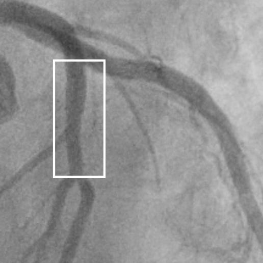
Given the news that his arteries had no plaque build-up, he felt no need to modify his lifestyle or take cholesterol medications in order to avoid the fate of early death from myocardial infarction that his father had suffered at age 50 and his sister at age 54.
Limitations Of The Cardiac Cath In Identifying Atherosclerotic Plaque
When I first saw him a year after the cardiac cath, I told him that although his previous cardiologist had told him all was fine with his coronary arteries he could, in fact, have significant diffuse subclinical atherosclerosis and still be at high risk for a heart attack.
This came as quite a shock to Jerry as he, like most laypeople, viewed the cardiac cath as the “gold standard” for assessment of the coronary arteries.
For most patients, a normal cath has been viewed as a warranty against heart attack
Although ICA has been the gold standard for the diagnosis of coronary artery disease, the lumenography it provides only shows the internal arterial lumen and does not reveal the vessel wall with its developing atherosclerotic plaque.
Previous studies analyzing serial angiograms from patients presenting with acute coronary syndrome (ACS) have suggested that in nearly two-thirds of the culprit lesions, the coronary angiogram obtained a few months before the acute event demonstrated no significant plaque impingement on the lumen.
Identifying Early Plaque Using Coronary Calcium Scans
I recommended the patient get a coronary artery calcium (CAC) scan to look for early coronary plaque and this demonstrated two small calcific plaques in the proximal portion of his LAD coronary artery (inside the green circle.)
His calcium score was 9 which is higher than 82% of 45-year-old white males.

Now that we had visual proof of the plaque in his arteries he was motivated to change his lifestyle to reduce the risk of suffering his dad’s fate. In addition, he was now willing to take medications to further reduce risk.
Best Imaging Test for the Patient With Symptoms Possibly Related to Coronary Disease
